Left Hip Muscles Anatomy : The Posterior Sling - Spontaneous Muscle Release ... / If you know all the hip flexor names and bones they attach to, that's an awesome accomplishment!
Left Hip Muscles Anatomy : The Posterior Sling - Spontaneous Muscle Release ... / If you know all the hip flexor names and bones they attach to, that's an awesome accomplishment!. The iliopsoas muscle is a major hip flexor. Learn their anatomy efficiently and easily using kenhub's muscle anatomy and reference charts! If left unstretched, shortened hip flexors affect the position of the pelvis, which in turn affects the position and movement of the lower back. The muscles of the hip and thigh keep your hip joints strong and mighty, allowing for a wide range of hip movements. Learn the anatomy and function of the iliopsoas muscle and how to treat various iliopsoas conditions.
This 20 x 26 (51 x 66 cm) wall poster shows location of various joints and provides anterior and posterior views of the left shoulder, right hip, right knee and left elbow. The cavity of the acetabulum the external obturator muscle is short external rotator muscle of hip joint. Most modern anatomists define 17 of these muscles, although some additional muscles may sometimes be considered. This anatomical atlas was especially designed for a specific public (radiologists, surgeons, rheumatologists and physicians specializing in musculoskeletal imaging). Most modern anatomists define 17 of these muscles, although some additional muscles may sometimes be considered.

3 months later i got acute excrutiating pain in inguinal area.
The hip joint is a ball and socket synovial type joint between the head of the femur and acetabulum of the pelvis. Most modern anatomists define 17 of these muscles, although some additional muscles may sometimes be considered. Learn their anatomy efficiently and easily using kenhub's muscle anatomy and reference charts! Knee assessment and hip mechanics learn how hip. Movement of the femur on the hip in a direction away from the midline of the body in the frontal plane. Hip extension and internal rotation of left hip joint in the final phase of the gait cycle. The main muscles of the hip and pelvis consistsof the iliopsoas, pectinues, rectus femoris and sartorius at the front. A bursa that sometimes causes problems in the hip is sandwiched between the bump on the outer hip (the greater trochanter) and the muscles and tendons that cross over the bump. Muscles, connected to bones or internal organs and blood vessels, are in charge for movement. Discover the muscle anatomy of every muscle group in the human body. In human anatomy, the muscles of the hip joint are those muscles that cause movement in the hip. Leave a comment cancel reply. Your email address will not be published.
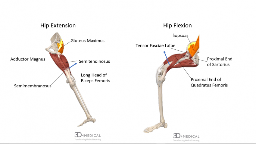
Advanced hip flexor muscle anatomy. Now that you watched the video, you. Learn their anatomy efficiently and easily using kenhub's muscle anatomy and reference charts! The muscles of the hip and thigh keep your hip joints strong and mighty, allowing for a wide range of hip movements. These muscles are responsible for hip joint extension (backward movement).

It is ideal for classrooms or doctor's offices, and.
The hip muscles encompass many muscles of the hip and thigh whose main function is to act on the thigh at the hip joint and stabilize the pelvis. These muscles work together to flex your hip and to stabilize your hip and lower back during activities such as walking, running, and rising from a chair. Anatomy of the muscular system. What is collectively referred to as the hip flexors is actually a group of muscles that includes the iliopsoas, the thigh muscles (rectus femoris, sartorius and tensor fasciae. In conclusion, a thorough understanding of pelvic and hip anatomy is important for. Their main function is contractibility. The geometry of the hip allows wide range of motion in all planes. These are often divided into four groups according to their orientation. It is a flat, triangular muscle on the anterior wall of the pelvis. Related online courses on physioplus. Movement of the femur on the hip in a direction away from the midline of the body in the frontal plane. Each muscle below has the bones in bold for intermediate learners and the specific bony landmarks for advanced learners. The muscles of the pelvis, hip and buttock anatomical chart shows how each muscle in this area of the body works with the others, and the you will not find a more comprehensive or more detailed examination of these muscles in an anatomy chart.
for detailed anatomy of pelvic bones, read anatomy of hip bone. Knee assessment and hip mechanics online course: The main muscles of the hip and pelvis consistsof the iliopsoas, pectinues, rectus femoris and sartorius at the front. Most modern anatomists define 17 of these muscles, although some additional muscles may sometimes be considered. It is the long, flat muscle that extends vertically between the pubis and both do side bending to the same side, but the external oblique on the left rotates the trunk/spine to the right, whereas the internal oblique on the left.
Your email address will not be published.
Each muscle below has the bones in bold for intermediate learners and the specific bony landmarks for advanced learners. Their main function is contractibility. Semimembranosus, semitendinosus and biceps femoris (the hamstrings). for detailed anatomy of pelvic bones, read anatomy of hip bone. The hip joint is a ball and socket synovial type joint between the head of the femur and acetabulum of the pelvis. Related online courses on physioplus. Leave a reply cancel reply. The iliopsoas muscle is a major hip flexor. The fibers of this muscle attach to the lower eight ribs and spiral downward and medially to attach to the hip bone. Knee assessment and hip mechanics learn how hip. Learn their anatomy efficiently and easily using kenhub's muscle anatomy and reference charts! Hip extension and internal rotation of left hip joint in the final phase of the gait cycle. What is collectively referred to as the hip flexors is actually a group of muscles that includes the iliopsoas, the thigh muscles (rectus femoris, sartorius and tensor fasciae.


Komentar
Posting Komentar